Busan Shoulder & Joint Surgery · Dedicated International Patient Care


Shoulder Joint Diseases · Needs Clinic Confirmation
Calcific Tendinitis
Calcific tendinitis occurs when calcium deposits build up within one of the rotator cuff tendons, most often the supraspinatus, causing pain that can range from mild to sudden and severe.
Schedule a ConsultationWhat Is Calcific Tendinitis? Understanding Calcium Deposits in the Shoulder

Calcific tendinitis occurs when calcium deposits build up within one of the rotator cuff tendons, most often the supraspinatus, causing pain that can range from mild to sudden and severe. Because the shoulder relies on the rotator cuff for its exceptional range of motion, even small calcium deposits can trigger significant discomfort, particularly during overhead movement or at night.
Calcific tendinitis treatment typically begins conservatively and escalates only as needed. Options include medication, injection therapy, ultrasound-guided needle aspiration (sometimes called barbotage) to break up and remove calcium deposits, and extracorporeal shock wave therapy (ESWT), which uses targeted sound waves to help fragment calcium buildup. Physical and manual therapy support the healing process throughout, with arthroscopic surgical removal reserved for deposits that do not respond to these non-surgical approaches. Since calcific tendinitis symptoms can resemble rotator cuff tear or shoulder impingement, accurate imaging-based diagnosis is an essential first step.
Who Needs Calcific Tendinitis Treatment?
Calcific tendinitis often develops without an obvious triggering injury, and candidates for evaluation typically share certain patterns.
- Adults in their 30s–50s experiencing shoulder pain without a clear history of trauma or overuse
- Patients who describe a sudden, sharp increase in shoulder pain, sometimes overnight
- Individuals with pain that intensifies during overhead reaching or specific arm movements
- Patients who have had shoulder imaging show calcium deposits within the rotator cuff
- Those experiencing night pain that disrupts sleep, particularly when lying on the affected shoulder
- Patients who have tried rest or anti-inflammatory medication without significant relief
- Individuals interested in non-surgical options such as ESWT or needle aspiration before considering surgery
- Patients with recurring shoulder pain episodes who want an accurate diagnosis to confirm the underlying cause
Meet the Shoulder Specialists
 |
 |
 |
|---|---|---|
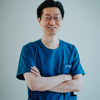
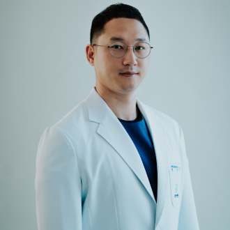
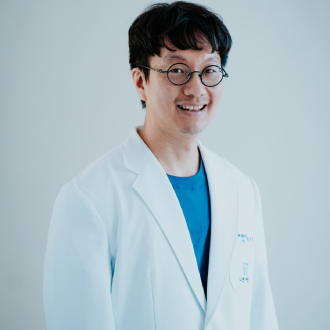
| Dr. Lee ChangsooOrthopedic Surgeon | Dr. Lee DongkiOrthopedic Surgeon | Dr. Rhim Chunsub Anesthesiologist |
Calcific tendinitis treatment at Narsha Hospital is provided by a team of fellowship-trained shoulder and elbow surgeons, supported by dedicated anesthesiology and imaging staff for cases requiring further intervention.
Dr. Lee Changsoo is a Professor of Shoulder Surgery at Inje University Paik Hospital who has also served as a Visiting Professor at Balgrist Hospital, University of Zurich, and the McKay Orthopaedic Research Laboratory at the University of Pennsylvania. He previously led the Joint Centers at Seoul Bumin Hospital and Haeundae Bumin Hospital, and is a member of both the Korean Orthopaedic Association and the Korean Shoulder and Elbow Society.
Dr. Lee Dongki is a Clinical Professor of Shoulder Surgery at Kyunghee University and a fellowship graduate of the Seoul Shoulder and Elbow Center, a program affiliated with Seoul National University, Samsung Hospital, and St. Mary's Hospital. His overseas training includes the Kerlan-Jobe Clinic, Keck Hospital at the University of Southern California, and Langone Hospital at New York University. His published research in the Journal of Shoulder & Elbow Surgery (JSES) reflects the diagnostic precision applied across the full range of shoulder conditions treated at the hospital, including calcific tendinitis.
Dr. Rhim Chunsub, an anesthesiologist trained at Busan National University, supports patients undergoing arthroscopic calcium deposit removal when non-surgical treatment has not resolved symptoms.
This team shares a care philosophy centered on confirming the diagnosis through imaging, prioritizing minimally invasive treatment where possible, and reserving surgery for cases that genuinely require it.
Calcific Tendinitis Treatment Areas and Expected Improvements
- Supraspinatus tendon: The most common site for calcium deposit formation in the shoulder. Treatment targeting this tendon, including ESWT or ultrasound-guided aspiration, aims to break down and reduce calcium buildup, helping to relieve the associated pain over time.
- Rotator cuff tendon sheath and surrounding soft tissue: Inflammation surrounding the calcium deposit often contributes significantly to pain intensity, particularly during the acute phase. Injection therapy may be used to help manage this localized inflammation.
- Subacromial space: Calcium deposits can sometimes contribute to a mechanical pinching sensation during overhead movement. Physical therapy techniques may help maintain shoulder mechanics and reduce discomfort during this stage of treatment.
- Surgical removal site (in resistant cases): For deposits that do not respond to conservative treatment, arthroscopic removal directly addresses the calcium buildup, aiming to relieve pain and restore more comfortable shoulder movement.
Most patients can expect a reduction in pain intensity and improved shoulder comfort as calcium deposits are addressed through treatment, though the timeline and degree of improvement vary based on deposit size, chronicity, and individual response. No specific outcome or recovery timeline can be guaranteed.
Calcific Tendinitis Treatment Process and Timeline
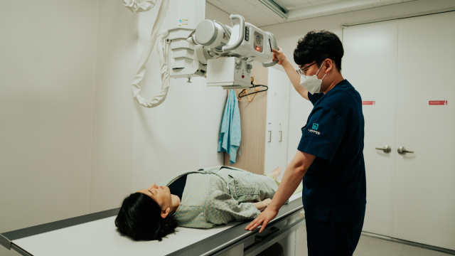
Treatment at Narsha Hospital follows a structured process designed to confirm the diagnosis and select the most appropriate, least invasive effective option.
- Initial Consultation and Imaging: A shoulder specialist evaluates symptoms, supported by in-house X-ray, MRI, or ultrasound to confirm the presence, size, and location of calcium deposits.
- Treatment Planning: Based on deposit characteristics and symptom severity, the care team determines whether ESWT, needle aspiration, injection therapy, or another approach is most appropriate.
- Non-Surgical Treatment: This may include ultrasound-guided needle aspiration to remove calcium deposits directly, ESWT sessions to help fragment remaining calcification, and physical therapy to support shoulder mechanics.
- Immediate Aftercare: Patients receive guidance on activity modification and pain management following aspiration or ESWT sessions.
- Reassessment: Follow-up imaging or evaluation tracks the reduction in calcium deposits and corresponding symptom improvement.
- Surgical Removal (if needed): For deposits resistant to conservative treatment, arthroscopic removal is performed in an ISO Class 5 (Class 100) cleanroom operating room, followed by structured rehabilitation.
Mild swelling, bruising, or soreness at the treatment site for a few days following needle aspiration or ESWT is common and generally resolves with standard aftercare guidance.
Calcific Tendinitis Results, Safety, and Recovery Considerations
Many patients notice symptom improvement within several weeks of beginning non-surgical calcific tendinitis treatment, particularly following needle aspiration or a course of ESWT sessions, though recovery timelines vary based on deposit size and individual response. Patients who require surgical removal typically follow a structured rehabilitation period afterward to support full recovery of shoulder function.
Non-surgical calcific tendinitis treatments generally carry a favorable safety profile, with potential side effects including temporary swelling, bruising, or soreness at the treatment site. For patients requiring arthroscopic removal, Narsha Hospital's ISO Class 5 cleanroom operating standards help minimize infection risk, though as with any procedure, complications are possible even if uncommon.
Results vary by individual, and no specific outcome or timeline can be guaranteed. Patients are encouraged to discuss realistic expectations for their specific presentation directly with their treating specialist.
Why Choose Narsha Hospital for Calcific Tendinitis Treatment?
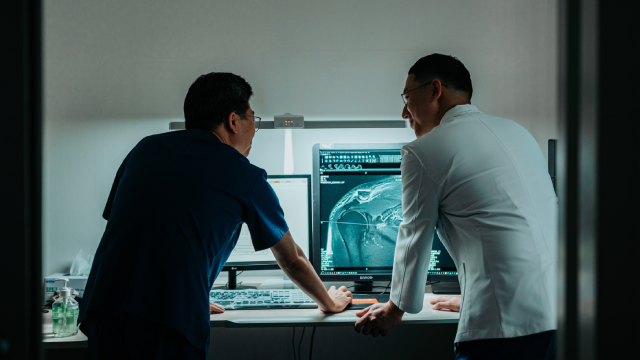
Narsha Hospital is one of the few hospitals in the region built exclusively around shoulder and elbow joint care, performing more than 1,200 shoulder and elbow surgeries annually across roughly 45,000 patient visits a year. This concentrated clinical focus supports precise identification and treatment of calcific tendinitis, distinguishing it clearly from other shoulder conditions with overlapping symptoms.
Diagnosis is supported by in-house X-ray, MRI, and ultrasound imaging, allowing for prompt confirmation of calcium deposit location and size without the delays of outsourced testing. For the smaller number of patients who require arthroscopic removal, procedures take place in operating rooms built to ISO Class 5 (Class 100) cleanroom standards with dedicated HEPA-filtered air handling. The hospital's overall treatment philosophy favors minimally invasive options such as ESWT and needle aspiration, reserving surgery for deposits that do not respond to these methods.
Clinic Highlights:
- Shoulder and elbow-focused specialist team led by Dr. Lee Changsoo and Dr. Lee Dongki, both fellowship-trained with international clinical experience
- In-house MRI, X-ray, and ultrasound imaging for precise calcium deposit localization
- ESWT and ultrasound-guided needle aspiration as first-line, minimally invasive treatment options
- Rehabilitation led by internationally certified physical therapists, supported by comprehensive nursing and caregiving services
The hospital's Narsha Shoulder & Joint Research Center continues to inform treatment protocols across all shoulder conditions, supported by the broader research contributions of its specialist team, including Dr. Lee Dongki's published work in JSES.
Real Patient Examples
Calcific Tendinitis Patient, Early 40s
A patient in their early 40s came in after experiencing a sudden, sharp increase in shoulder pain without any preceding injury. Imaging confirmed a calcium deposit within the rotator cuff, and the patient underwent ultrasound-guided needle aspiration combined with a short course of physical therapy. The patient reported meaningful pain relief within several weeks of treatment.
Calcific Tendinitis Patient, Late 30s
A patient in their late 30s experienced recurring shoulder pain episodes over several months, with imaging revealing a persistent calcium deposit. After a course of extracorporeal shock wave therapy sessions, the patient noted a gradual reduction in pain and improved comfort during overhead movement.
Take the Next Step Toward Shoulder Comfort
Sudden or recurring shoulder pain from calcium buildup doesn't have to be a long-term issue — an accurate diagnosis can help identify the most effective, least invasive treatment path for your condition.
[Schedule Your Calcific Tendinitis Evaluation] — Connect with Narsha Hospital's shoulder specialist team for an in-house imaging evaluation and a personalized discussion of your treatment options, from ESWT and needle aspiration to surgical removal when needed, guided by fellowship-trained surgeons with international clinical experience in shoulder and elbow medicine.
For questions or to speak with our appointment desk, call our patient coordination team, who can help schedule your consultation and answer initial questions about the evaluation process.
Other Services
Explore additional treatments offered by our specialists.

Digestive Disorders
Digestive disorders encompass a wide range of conditions affecting the esophagus, stomach, duodenum, and colon, often producing symptoms such as abdominal discomfort, bloating, changes in bowel habits, or difficulty digesting food.
Learn More
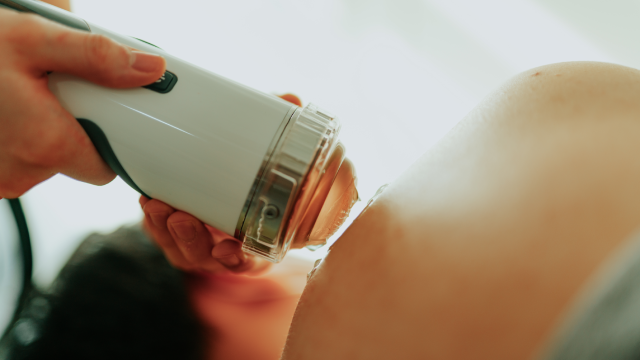
Non-surgical Sports Treatment
Non-surgical sports treatment refers to conservative care for shoulder and elbow injuries sustained during athletic competition, training, or recreational sports activity, without the need for an operation.
Learn More
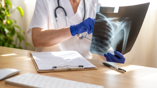
Shoulder Fracture
A shoulder fracture refers to a break in one of the bones that make up the shoulder girdle — most commonly the proximal humerus, but also the clavicle or scapula — typically resulting from a fall, direct impact, or high-energy trauma.
Learn More